Research Article | | Peer-Reviewed

Characterization of Fermentative Strains Isolated from Spontaneously Fermentation of Cashew Apples (Anacardium occidentale L.)

Received: 12 January 2026     Accepted: 26 January 2026     Published: 6 February 2026
Views:       Downloads:
Abstract

The cashew apple is a juicy and sweet fruit that loses its nutritional quality during the first week after falling from the cashew tree. This degradation is strongly linked to microorganisms that may present beneficial biotechnological potential. Therefore, the objective of this study is to characterize lactic acid bacteria and fermentative yeasts from cashew apples. Cashew apples collected from three localities (Tioro, Morovine, and Waraniene) in the Korhogo department (Northern Côte d’Ivoire) were washed in the laboratory, crushed, and subjected to fermentation. Samples were taken every two days over one week. Morphological studies focused on macroscopic and microscopic analyses of the strains. Physiological and biochemical tests of lactic acid bacteria and yeast isolates were all carried out in tubes. Molecular typing of these isolates was performed using the restriction enzymes DDeI or HinfI, and HaeIII. Characterization of lactic isolates revealed that all lactic acid bacteria isolated were bacillary in shape with catalase activity. They are homofermentative, mesophilic, acidophilic, and do not tolerate salinity beyond 4%. Moreover, they fermented lactose, glucose, maltose, fructose, starch, sucrose, and cellulose. They also produced exopolysaccharides. At the molecular level, all bands were observed at 1500 base pairs. Furthermore, the biochemical profile and enzymatic digestion performed with restriction enzymes identified the genus Lactobacillus, specifically Lactobacillus plantarum as the presumptive species. Characterization revealed yeasts of oval, elongated shape with lateral budding. Sediments were observed in their growth broths. They degraded urea and produced acetoin. They also fermented various sugars, except cellulose, citrate, and mannitol. All bands were observed at 500 base pairs. Biochemical tests and PCR confirmed that all isolates belonged to the genus Saccharomyces sp. Digestion with restriction enzymes (HaeIII and HinfI) divided them into two groups of Saccharomyces, of which 93.75% were Saccharomyces cerevisiae and 6.25% were Saccharomyces uvarum. The use of these presumptive species could serve in the production of cashew apple wine.

Published in International Journal of Microbiology and Biotechnology (Volume 11, Issue 1)
DOI 10.11648/j.ijmb.20261101.12
Page(s) 9-19
Creative Commons

This is an Open Access article, distributed under the terms of the Creative Commons Attribution 4.0 International License (http://creativecommons.org/licenses/by/4.0/), which permits unrestricted use, distribution and reproduction in any medium or format, provided the original work is properly cited.

Copyright

Copyright © The Author(s), 2026. Published by Science Publishing Group

Keywords

Cashew Apple, Fermentation, Lactic Acid Bacteria, Yeast, Enzymatic Digestion

1. Introduction
Since the Neolithic era, fermentation has been used by humans to preserve food and improve its organoleptic and nutritional qualities . The Sumerians, in Mesopotamia, were already practicing fermentation to produce alcoholic beverages 8000 years before Christ . It was not until the 18th century that the microorganisms involved in fermentation were scientifically identified . Since then, fermentation has been recognized as a major biotechnological tool. From a biochemical perspective, fermentation is the transformation of a substrate under the action of microorganisms or their enzymes . It can occur either spontaneously and naturally, or in a controlled manner that meets industrial requirements . Thus, fermentation is no longer perceived merely as a preservation method, but as a true platform for the production of high value-added biomolecules. From an industrial standpoint, fermentation refers to technological processes that enable microbial development and metabolite production, under aerobic or anaerobic conditions . The cashew tree is cultivated in northern Côte d’Ivoire mainly for its fruit, especially for its nut. Once detached from the tree, the cashew apple deteriorates very quickly. Its nutritional quality declines during the first week after detachment, which limits its use. The degradation of this fruit is strongly linked to the activity of a diversity of microorganisms that may present beneficial biotechnological potential. The microorganisms of greatest biotechnological interest used in fermentation processes, particularly in the agri-food sector, are lactic acid bacteria, acetic acid bacteria, and yeasts , corresponding to lactic, propionic, acetic, and alcoholic fermentations. Natural fermentation of cashew apples may contain a wide range of lactic acid bacteria and yeasts whose identities are not well known. These microorganisms could have particular biotechnological interest, potentially promoting the valorization of cashew apples produced in Côte d’Ivoire. For this reason, the biochemical and molecular characterization of lactic acid bacteria and yeasts involved in cashew apple fermentation was the focus of our study.
2. Materials and Methods
2.1. Plant Material
The biological material consisted of red and yellow cashew apples collected from three localities (Tioro, Morovine, and Waraniene) in the Korhogo department, Côte d’Ivoire. These cashew apples were harvested in March 2024 from four orchards in these localities.
2.2. Preparation of Cashew Apple Samples
Once harvested, the apples were carefully separated from the nuts, then transported to the laboratory where they were washed with distilled water. They were subsequently crushed using a blender (SMART model No.: STPE1110). One (1) kilogram of the crushed material of each color was collected and transferred into Erlenmeyer flasks, sealed with carded cotton and covered with aluminum foil (Figure 1). Fermentation was carried out at ambient temperature for six (6) days, with samples taken every two days during the fermentation process .
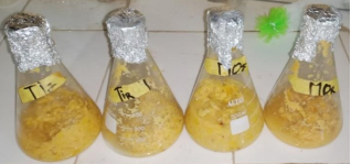
Figure 1. Crushed red and yellow cashew apples.
TiJ: yellow apple from Tioro
TiR: red apple from Tioro
MoJ: yellow apple from Morovine,
MoR: red apple from Morovine
2.3. Preparation of Culture Media
Isolation was carried out by surface streaking. For the isolation of lactic acid bacteria, 1% nystatin was added to the MRS medium after cooling, using a five (5) mL syringe. For the isolation of yeasts, 0.5% vancomycin and 0.5% gentamicin were added to the OGA medium (Oxytetracycline-Glucose-Yeast Extract Agar) after cooling . Once the media poured into Petri dishes had solidified, streak inoculation was performed directly using a platinum loop . The inoculated plates were then incubated at 30°C for 24 hours for yeasts and 48 hours for lactic acid bacteria .
2.4. Purification of Cashew Apple Isolates
The purification of strains was carried out through successive subculturing: on MRS agar for lactic acid bacteria and on PCA agar for yeasts. This consisted of re-streaking colonies onto Petri dishes containing the appropriate solid media, followed by incubation under temperature and duration conditions specific to each strain. This operation was repeated until pure colonies were obtained .
2.5. Identification of Strains
After obtaining homogeneous cultures, morphological, physiological, biochemical, and molecular tests were performed .
2.5.1. Morphological Study
The morphological study was based on macroscopic and microscopic observations . It was carried out on pure colonies.
Macroscopic observation highlighted the size, color, and shape of the colonies .
Microscopic observation allowed visualization of the shape and arrangement of cells as well as Gram type (for lactic acid bacteria). To achieve this, a smear was prepared on a slide from a young bacterial suspension. The slide was successively treated with gentian violet, Lugol’s iodine, alcohol, and fuchsin for 30 seconds each, with washing, rinsing, and drying between the different staining steps.
For yeasts, a sample from each pure strain was taken to determine the shape of the constitutive cells (spherical or oval), cell size, mode of reproduction, and the position of the bud(s) on the mother cell . The study consisted of examination under a light microscope at ×100 magnification, after Gram staining for bacteria and simple staining for yeasts.
2.5.2. Study of Physiological Tests of Lactic Acid Bacteria and Yeasts
The catalase test was performed by bringing a bacterial colony into contact with hydrogen peroxide. Gas release indicated positive catalase activity.
Growth at different temperatures was tested at 10°C, 37°C, and 45°C in tubes containing MRS broth. The tubes were examined after 24 hours, and growth was assessed by the appearance of turbidity .
Growth at different pH values was tested in MRS broths adjusted to pH 9.6, 5, 3.8, and 3 using NaOH and HCl . Growth was indicated by turbidity in the broth tubes.
The thermoresistance test of the strains was performed at 60°C for 30 minutes in MRS broth in a water bath, using a young and pure bacterial culture .
Growth in saline medium was tested in MRS broth containing 4% and 6.5% NaCl. The tubes were incubated at 37°C for 24 hours. Lactic streptococci are unable to survive in this medium .
2.5.3. Biochemical Study
Growth of lactic isolates in Sherman’s milk was tested on skimmed milk containing 0.1% and 0.3% methylene blue .
The search for arginine dihydrolase activity was carried out by adding a few drops of arginine to 1 ml of young culture. After incubation at 37°C for 24 hours, arginine-positive strains prevented the color change to yellow .
Acetoin production was tested using the Voges-Proskauer (VP) reaction, applied as follows: in a test tube containing 1 ml of the culture to be tested, 5 drops of reagent VPI (16% NaOH solution in distilled water) and 5 drops of reagent VPII (6% α-naphthol in 95% ethanol) were added. After resting for 5 to 10 minutes at room temperature, a positive test was indicated by the appearance of a pink ring on the surface of the medium .
The fermentative type of lactic isolates was determined by introducing Durham tubes into broth tubes. Gas release by heterofermentative bacteria accumulated in the Durham tube after incubation at 37°C for 24 to 48 hours .
Dextran production from sucrose was demonstrated on liquid and solid Mayeau, Sandine, and Elliker (MSE) medium . Dextran-producing strains were characterized by the formation of large, viscous, and slimy colonies on Petri dishes.
The sugars used were: hexoses (D-glucose, D-fructose), disaccharides (lactose, maltose, sucrose), and polysaccharides (starch, cellulose). The sugars used as carbon sources were prepared at 2%. After 24 to 48 hours of incubation at 37°C, a color change to yellow indicated sugar utilization by bacteria or yeasts .
Urease activity was detected on Christensen medium. A positive reaction was indicated by the appearance of a bright pink to red coloration in the culture medium after 3 days of incubation at 30°C.
Simmons citrate was used as a carbon source, with central streak inoculation performed along the slope of the tubes, from the bottom to the surface of the medium. Color change was observed after 24 hours of incubation at 30°C.
On mannitol-motility medium, inoculation was performed by central stabbing, stopping 1 cm from the bottom of the tube. After 24 hours of incubation at 30°C, color change and turbidity of the medium were noted.
For the coagulation test, lactic isolates were inoculated into flasks containing sterile skimmed milk reconstituted at 10%. Observation was carried out after 24 hours of incubation at 37°C .
2.5.4. Molecular Study of Lactic Acid Bacteria and Yeasts
DNA extraction from the strains was carried out following the protocol described by El Sheikha et al. . Strains were collected from Petri dishes under aseptic conditions and introduced into 2 mL Eppendorf tubes (in duplicate) using sterile distilled water with 0.1% Tween 80. Glass beads (0.3 g) with a diameter of 425–600 µm (Sigma, France) were added to the tube contents. The mixture was vigorously shaken for 30 minutes in a Vortex mixer and then centrifuged at 8000 g for 15 minutes to remove the supernatant. The cell pellet was resuspended in a solution composed of 300 µL hydrolysis buffer, 100 µL TE (10 mM Tris-HCl, 1 mM EDTA, pH 8.0), 100 µL lysozyme solution, and 100 µL proteinase K solution. The mixture was incubated at 42°C for 20 minutes. Then, 50 µL of 20% SDS were added to each tube, followed by incubation at 42°C for 10 minutes. In addition, 400 µL of MATAB were added to each tube and incubated at 65°C for 10 minutes after vigorous shaking for 5 minutes. Furthermore, 700 µL of phenol/chloroform/isoamyl alcohol (25/24/1, v/v/v) were added twice to the tube contents, shaken in a vortex for 5 minutes, and centrifuged again at 8000 g for 15 minutes. The aqueous phase was transferred to an Eppendorf tube, and residual phenol was removed using 600 µL chloroform/isoamyl alcohol (24/1) after centrifugation at 8000 g for 15 minutes. The aqueous phase was collected, and DNA was stabilized in 30 µL sodium acetate (3 M, pH 5), followed by precipitation with an equal volume of chilled isopropanol. DNA in isopropanol was stored at –20°C for 12 hours (overnight). After centrifugation at 8000 g for 15 minutes, the supernatant was discarded, and the DNA pellets obtained were washed with 500 µL of 70% ethanol. Ethanol was removed after centrifugation. Finally, DNA was resuspended in 50 µL ultrapure water and stored at –20°C until analysis.
Composition of the PCR reaction mixture was performed in a reaction volume containing 5 µL template DNA, 5 µL sample DNA, 1 µL of each primer, 1 µL deoxynucleoside triphosphates (dNTP, 2.5 mM, brand D0056, Sangon Biotech., Shanghai, Co., Ltd., China), and 20 µL DNA polymerase (DyNAzyme EXT) . The universal 16S DNA primers for bacteria used were:
1) 7F 1540R (5-AGA GTT TGA TCC TGG CTC AGG AGG TGA TCC AGC CGC A-3)
2) 1492R (5-AGT TTGATCMTGGCTCAG GGT TAC CTT GTT ACG ACT T-3)
These amplify fragments of 1500 bp .
The 26S DNA primers for yeasts used were:
1) NL1 (5-GCATATCAATAAGCGGAGGAAAAG-3)
2) NL4 (5-GGTCCGTGTTTCAAGACGG-3)
These amplify fragments of 500 bp .
Amplification of lactic acid bacteria and yeast genes was carried out using a PCR thermal cycler (2720 thermal cycler, Applied Biosystems) under the conditions shown in the following Table 1. Finally, the amplicons were stored at 4°C .
Table 1. Gene Amplification Program.

Amplification program

Lactic acid bacteria

Yeasts

Initial denaturation

95°C / 4 min

95°C/3 min

Denaturation

95°C / 60 s

95°C / 30 s

Annealing

57°C / 60 s

55°C / 60 s

Extension

72°C / 1 min

72°C / 1 min

Final extension

72°C / 5 min

72°C / 10 min

Gel electrophoresis migration, agarose (1.2% w/v) was dissolved by heating in 1X TAE buffer. An intercalating DNA agent, ethidium bromide (EtBr), was then added at a final concentration of 0.2 µg/mL. After polymerization, the gel was placed in an electrophoresis tank containing 1X TAE buffer. Subsequently, 4 µL of each prepared DNA sample was mixed with 1 µL of 10% (v/v) loading dye and loaded into the wells. Electrophoresis was performed at 90 V for 45 min for lactic acid bacteria and at 150 V for 20 min for yeasts. DNA bands were visualized under UV light using an Infinity Capt imaging system (Fisher Scientific, Bioblock). The presence of bands corresponding to the amplified fragments was compared with the molecular size marker .
Composition of the reaction mixture for gene digestion, PCR-amplified DNA fragments were purified using the EZ-10 Spin Column PCR DNA Extraction Kit. The reaction mixture for restriction digestion of bacterial and yeast genes consisted of 12.3 µL of double-distilled water, 2 µL of 10X RE buffer, 0.2 µL of acetylated SAB (10 µg/µL), 0.5 µL of each restriction enzyme (DDeI, HaeIII, and HinfI), and 5 µL of DNA from each sample. All digestion reactions were incubated at 37°C for 4 h. Enzymatic reactions were terminated by incubation at 65°C for 20 min to inactivate the enzymes .
3. Results
Inoculation allowed the isolation of pure colonies of lactic acid bacteria and yeasts. Consequently, three (03) lactic acid bacterial isolates and three (03) yeast isolates were selected during sampling. From 24 samplings obtained from six (06) samples, a total of 144 isolates were recovered, including 72 lactic acid bacteria and 72 yeasts (36 isolates from red cashew apples and 36 from yellow cashew apples) during the fermentation of cashew apple pulp. For subsequent analyses, 16 lactic acid bacterial isolates and 16 yeast isolates were retained after morphological and physiological tests.
3.1. Characteristics of Fermentative Lactic Acid Bacterial Isolates from Cashew Apples
Macroscopic analysis of the cultivated bacterial colonies revealed circular, rounded, slightly convex, white colonies with a creamy and shiny appearance and a homogeneous aspect on MRS agar (Figure 2a). Gram staining showed that all isolates were Gram-positive. They were sometimes arranged in chains, as diplobacilli, or as single cells (Figure 2b).
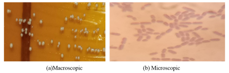
Figure 2. Appearance of Lactic Acid Bacteria.
All isolates were catalase-negative (−) and urease-negative (−). They did not grow at pH 3 or at pH 9.6. The isolates survived at 60°C for 30 minutes but did not grow at 10°C or 45°C. All isolates grew at 37°C. This characterization revealed that all isolates were acidophilic, thermoresistant, mesophilic, and tolerant only to low salinity (4%).
The biochemical study indicated that all lactic acid bacterial isolates were homofermentative, with positive results for the fermentation of skimmed milk, glucose, sucrose, fructose, lactose, maltose, and starch. Citrate, methylene blue (Sherman milk), ADH, and mannitol were not degraded. The isolates produced exopolysaccharides but were negative for acetoin production.
Genotype analysis showed that all lactic acid bacterial isolates aligned at 1500 base pairs on the electrophoresis gel (Figure 3a). The letter M represents the molecular weight marker. Digestion with the restriction enzymes DDeI and HaeIII revealed identical fragment patterns for all lactic acid bacterial isolates on the electrophoresis gel (Figure 3b and 3c). Furthermore, the sum of the fragment sizes generated by DDeI and HaeIII digestion was identical both to each other (Figure 3b and 3c) and to the 16S rDNA gene size (Figure 3a). This resulted in the formation of a single group of lactic acid bacteria after enzymatic digestion. This lactic acid bacterial group is therefore likely the dominant lactic species involved in cashew apple fermentation.
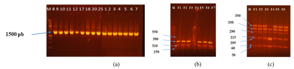
Figure 3. Electrophoretic profile of the 16S gene and restriction digestion profiles of lactic acid bacterial genes using DDeI and HaeIII.
(a) Electrophoretic profile of the 16S gene of lactic acid bacteria
(b) Digestion profile using DDeI
(c) Digestion profile using HaeIII
Lactic acid bacterial isolates: F1, F2, F3, F4, F5, F6, F7, F8, F9, F11, F12, F17, F18, F20 and F25
3.2. Characteristics of Yeast Isolates Obtained During Cashew Apple Fermentation
All isolated yeasts exhibited a white color with a regular circular margin, smooth surface, and convex elevation, and were oval in shape (Figure 4a). Growth of the yeasts in YPD broth showed the absence of a surface film and the presence of a white sediment (pellet) at the bottom of the tubes after 24 h of incubation at 30°C, with the exception of yeast Z.
Microscopic examination using simple staining on slides at 100× magnification revealed yeasts of varying sizes (elongated and short oval forms) with lateral budding (Figure 4b).
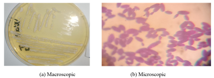
Figure 4. Appearance of yeasts.
All physiological tests performed on the yeast isolates yielded positive results, except in media with pH values of 3 and 9.6. Biochemical test indicated that the yeasts showed variable fermentation of Simmons citrate medium and nitrate–mannitol medium. Furthermore, all yeasts produced acetoin and fermented glucose, sucrose, fructose, lactose, maltose, and starch, except cellulose.
The electrophoretic profile showed that all isolated yeasts exhibited a similarity of 500 base pairs on the electrophoresis gel (Figure 5a).
The letter M represents the molecular weight marker. Digestion with the restriction enzymes HinfI and HaeIII revealed identical fragment patterns for all yeasts on the electrophoresis gel, except for yeast Z (Figure 5b and 5c). The restriction enzymes allowed the grouping of the isolated yeasts into two distinct groups (Figure 5b and 5c). However, the sizes of the different genomic fragments generated by HinfI and HaeIII digestion differed between the enzymes. The molecular size difference observed between yeast Z and the other yeasts enabled their classification into two groups after enzymatic digestion.
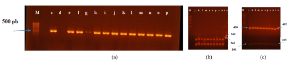
Figure 5. Electrophoretic profile of the 26S gene (a) and restriction digestion profiles of yeast genes using the enzymes HinfI (b) and HaeIII (c).
(a) Electrophoretic profile of the 26S gene of yeasts
(b) Digestion profile using HinfI
(c) Digestion profile using HaeIII
Yeast isolates: c, e, c, f, h, i, j, k, l, m, n, o, p, s, u, v, and z
4. Discussion
In this section, authors are advised to provide a thorough analysis of the results and make comparisons with relevant literature, not a short summary or conclusion. Any future research directions could also be stated in the discussion. Morphological analyses revealed that the dominant microflora during cashew apple fermentation consisted of rod-shaped lactic acid bacteria. The observed characteristics correspond to the classical descriptions of fermentative genera most frequently encountered in tropical fruits and fruit juices. The homogeneity of colony morphology highlights a relatively stable fermentative environment largely dominated by lactic acid bacteria. This predominance can be explained by the natural richness of cashew apples in fermentable sugars (glucose and fructose), which favor their development . The complete absence of cocci suggests that fermentation was mainly carried out by species of the genus Lactobacillus. This microflora, typical of spontaneous fermentations of tropical fruits, plays an essential role in stabilization, acidification, and nutritional valorization of the product . These observations are consistent with literature data on tropical fruit fermentations (pineapple, mango, cashew apple), where Lactobacillus plantarum and Lactobacillus fermentum are generally the dominant species . Physiological tests are essential criteria for the classification of lactic acid bacteria (LAB). The growth of lactic isolates between pH 3.8 and 6.5 allows effective fermentation of cashew apple juice or pulp, thereby reducing the pH and making the environment unfavorable for the development of pathogenic microorganisms . Moderate osmotic resistance at approximately 4% NaCl enables their survival in environments rich in fermentable sugars, typical of tropical fruits. Thermoresistance and growth at 37°C demonstrated optimal adaptation of these isolates to fermentation conditions in tropical regions, while remaining sensitive to extreme temperatures and harsh storage conditions. These characteristics are consistent with reports on lactic acid bacteria involved in the fermentation of tropical fruits such as cashew apple, pineapple, and mango . These studies highlight the dominance of this lactic genus during spontaneous fermentation and its key role in stabilization and acidification of the final product. The uniformity of morphological and physiological test results indicates a homogeneous dominant microflora, likely composed of species well adapted to pulp fermentation. These findings corroborate those of Bekhouche and Mechai , who confirmed that most Lactobacillus species thrive in acidic environments and do not tolerate salinity above 5%. Kamar reported that among the collected isolates, only Lactobacillus plantarum was unable to grow at 45°C, which is consistent with the results obtained in the present study on lactic acid bacteria from cashew apple fermentation. The biochemical profile highlighted the functional role of lactic acid bacteria during the fermentation process. The absence of CO₂ production in Durham tubes and the lack of acetoin production suggest that the isolates are homofermentative, producing mainly lactic acid as the final product of sugar metabolism, which is ideal for product acidification. The ability of the isolates to ferment various sugars indicates their adaptation to fermentable sugar-rich substrates, enabling efficient fermentation of cashew pulp and contributing to the formation of organic acids responsible for flavor and preservation . The degradation of starch and cellulose demonstrates their role in converting polysaccharides into simple sugars, thereby facilitating the release of fermentable sugars and accelerating spontaneous fermentation . The production of exopolysaccharides (EPS) improves texture, viscosity, and microbial stability of fermented juices or pulps while providing potential protective effects against environmental stress . None of the isolates produced catalase or urease, nor degraded ADH or citrate. Meryem reported that citrate lyase enzymes are present in strains belonging to the genera Streptococcus, Lactococcus, and Enterococcus. These findings are supported by Houali , who stated that Lactobacillus species do not possess citrate lyase capable of degrading citrate to produce acetoin. Similarly, studies by Bourgeois et al. and Leveau et al. showed that citrate and acetoin tests are useful for identifying Streptococcus, Lactococcus, Pediococcus, and Enterococcus. The results are consistent with those reported by Bouchibane in Algeria and Morocco during LAB characterization, where Lactobacillus casei subsp. casei, Lactobacillus casei subsp. alactosus, Lactobacillus plantarum, and Lactobacillus amylophilus degraded the same substrates. Huang et al. demonstrated that Lactobacillus plantarum and Lactobacillus fermentum are generally the dominant species in tropical fruit fermentations (pineapple, mango, cashew apple). Moreover, Lactobacillus fermentum is a facultative anaerobe, unlike Lactobacillus plantarum , which supports the identification of Lactobacillus plantarum as the presumptive species. The morphological, physiological, and biochemical profiles of these isolates are highly similar, reinforcing the hypothesis that Lactobacillus plantarum is the dominant species. These biochemical characteristics confirm the fundamental functional role of Lactobacillus plantarum in cashew apple fermentation, in agreement with previous studies on tropical fruit fermentations. Genomic analysis revealed that all lactic isolates exhibited amplicons of approximately 1500 base pairs, confirming their identity as Lactobacillus species, in addition to the use of MRS selective medium and microscopic examination. Genotypic methods such as PCR and restriction fragment length polymorphism (PCR-RFLP) were used for isolate typing. All isolates showed identical 16S gene digestion patterns, indicating strong genetic similarity. Restriction enzymes (DDeI and HaeIII) generated multiple fragments with identical base pair sizes for each isolate. Each band was associated with a specific base pair size according to its migration on the electrophoresis gel. These results indicate that all isolates belong to a homogeneous restriction profile, suggesting that they represent the same species. This technique is widely used in microbiology for typing lactic acid bacteria and confirming species-level identity . This confirms that the presumptive dominant species is L. plantarum, consistent with previous morphological, physiological, and biochemical analyses. Digestion with HaeIII revealed seven fragments of the following sizes for all isolates: 50 bp, 60 bp, 205 bp, 215 bp, 290 bp, 330 bp, and 350 bp. Aspasia et al. obtained seven fragments for Lactobacillus plantarum using the MseI restriction enzyme, with fragment sizes (90 bp, 110 bp, 140 bp, 160 bp, 270 bp, 290 bp, and 480 bp) totaling approximately 1500 bp. Similar results were observed for 16S rDNA digestion in the present study. Differences in fragment sizes may be attributed to the restriction enzymes used in PCR-RFLP. Digestion with DDeI produced four fragments (250 bp, 310 bp, 390 bp, and 550 bp), totaling approximately 1500 bp for all isolates. Thus, lactic fermentation of cashew apples was dominated by Lactobacillus plantarum. Differences in fragment number and size are explained by the distinct restriction sites specific to each enzyme.
The characteristics of yeast colonies white color, smooth surface, convex margin, oval shape, and budding reproduction correspond to the profiles of many fermentative yeasts, particularly Saccharomyces spp., Candida spp., or Pichia spp. . Lateral budding is characteristic of yeasts belonging to the family Saccharomycetaceae and constitutes a preliminary identification criterion . The absence of surface film observed in all isolates indicates that these yeasts do not form pellicles, unlike Pichia or Candida species . Consequently, the genus Saccharomyces is most likely involved. Cashew apples appear to be colonized mainly by a group of morphologically identical yeasts, possibly due to their high sugar content. The presence of white, oval, budding, and readily sedimenting yeasts is typical of yeasts used in alcoholic fermentation . All yeast isolates shared very similar morphological characteristics, suggesting a uniform yeast population likely dominated by a single species. Physiological tests provided insight into the metabolic behavior, enzymatic activity, and resistance of yeast isolates from cashew apple fermentation. All yeast isolates exhibited the same enzymatic profile. Catalase activity is common among fermentative yeasts and promotes survival in acidic, sugar-rich environments. Catalase positivity indicates the ability to degrade hydrogen peroxide, allowing adaptation to oxidative environments generated during fermentation. The isolates tolerated hyperosmotic conditions, a characteristic of yeasts originating from sugary fruits. Optimal growth of fermentative yeasts generally occurs between pH 3 and 6, confirming their good adaptation to cashew apple fermentation . Yeasts isolated from cashew apples exhibited a metabolic profile enabling utilization of not only simple sugars (glucose, fructose, sucrose) but also polysaccharides such as starch. The ability of all isolates to ferment glucose, fructose, maltose, and sucrose indicates a biochemical profile clearly associated with species of the genus Saccharomyces, particularly Saccharomyces. cerevisiae . Partial reactions with mannitol and lactose suggest poor assimilation of polyols such as mannitol . These yeasts metabolize lactose poorly or not at all, a characteristic of Saccharomyces species . Biochemical profiles indicated that 15 out of 16 isolates corresponded to Saccharomyces cerevisiae, the most common yeast in fruit fermentations. The ability of these yeasts to ferment multiple sugars suggests their potential application in bioethanol production. Acetoin production by these isolates may also be of interest to the food and fermented beverage industries. The 26S rDNA (NL region) of yeasts isolated from cashew apples was digested using two restriction endonucleases (HinfI and HaeIII). Two groups of isolates were subsequently defined based on cleavage profiles. Restriction products generated by HinfI and HaeIII indicated that the isolates belonged to two presumptive Saccharomyces species. Yeast Z alone constituted the second group, representing a distinct species due to its different migration pattern. Enzymatic digestion results were consistent with biochemical test outcomes. Antonio et al. reported the presence of two fragments produced by HinfI digestion in Saccharomyces cerevisiae strains, with PCR-RFLP profiles identical to those observed in the present study. Furthermore, digestion with HaeIII generated fragments of approximately 520 bp (110 bp + 480 bp), which are generally similar to those described by Andrea et al. . These authors obtained fragments of 480 bp (180 bp + 300 bp) with HaeIII and a single fragment of 770 bp with EcoRI for Saccharomyces uvarum. For Saccharomyces cerevisiae, they observed a 480 bp fragment with HaeIII and two fragments of 770 bp (500 bp + 270 bp) following EcoRI digestion. Variations in fragment sizes depending on the enzymes used may be explained by the specificity of restriction sites unique to each enzyme.
5. Conclusion
This part of the doctoral research, which falls within the framework of identifying lactic acid bacteria and yeasts involved in the fermentation of cashew apples, led us to perform morphological, physiological, biochemical, and molecular characterizations. Regarding lactic acid bacteria, morphological, physiological, and biochemical identification showed that Lactobacillus plantarum was the only species present. Similarly, molecular identification and enzymatic digestion using the restriction enzymes DDeI and HaeIII confirmed the presence of a single group of Lactobacillus plantarum. With respect to yeasts, morphological, physio-logical, molecular, and biochemical identification indicated that the yeasts responsible for alcoholic fermentation of cashew apples belong to the genus Saccharomyces. Restriction analysis of isolate genes using the enzymes HinfI and HaeIII allowed the classification of Saccharomyces isolates into two distinct groups.
Abbreviations

TiJ

Yellow Apple from Tioro

TiR

Red Apple from Tioro

MoJ

Yellow Apple from Morovine

MoR

Red Apple from Morovine

PCR

Polymerase Chain Reaction

PCA

Plate Count Agar

OGA

Oxytetracycline-Glucose-Agar

MRS

De Man, Rogosa, and Sharpe

VP

Voges-Proskauer

dNTP

Deoxynucleoside Triphosphates

RFLP

Restriction Fragment Length Polymorphism

YPD

Yeast Peptone Dextrose

EDTA

Ethylenediaminetetraacetic Acid

SDS

Sodium Dodecyl Sulfate

DNA

Deoxyribonucleic Acid

ADH

Arginine Dihydrolase Activity

EtBr

Ethidium Bromide

bp

Base Pairs

Acknowledgments
The authors would like to express their sincere and profound appreciation to the staff of the Laboratory of Biotechnology and Valorization of Agro-resources and Natural Substances at Peleforo GON COULIBALY University (Korhogo, Côte d’Ivoire), the Microbiology Laboratory of the Yopougon Technical High School (Abidjan, Côte d’Ivoire), and the Laboratory of Microbiology and Molecular Biology at Nangui ABROGOUA University (Abidjan, Côte d’Ivoire) for their invaluable collaboration and support.
Author Contributions
Bazoumana Fofana: Conceptualization, Investigation, Methodology, Writing – original draft, Writing – review & editing
Armel Fabrice Zoro: Supervision, Writing – review & editing
Abdoulaye Toure: Investigation, Methodology, Validation, Writing – review & editing
Tidiane Kamagate: Methodology, Supervision, Writing – original draft, Writing –review & editing
Safiatou Traore: Methodology, Writing – review & editing
Yade Rene Soro: Methodology, Writing – review & editing
Data Availability Statement
The data supporting the outcome of this research work has been reported in this manuscript.
Conflicts of Interest
The authors declare no conflicts of interest.
References
[1] Kalui, C. M., Mathara, J. M., Kutima, P. M. Probiotic potential of spontaneously fermented cereal-based foods – a review. African Journal of Biotechnology; 2010, 9(17), 2490–2498.
[2] Bourdichon, F., Casaregola, S., Farrokh, C., Frisvad, J. C., Gerds, M. L., Hammes, W. P., Harnett, J., Huys, G., Laulund, S., Ouwehand, A., Powell, I. B., Prajapati, J. B., Seto, Y. T., Schure, E., Van Boven, A., Vankerckhoven, V., Zgoda, A., Tuijtelaars, S., Hansen, E. B. Food fermentations: Microorganisms with technologically beneficial use. International Journal of Food Microbiology; 2012, 154, 87–97.
[3] ASTEP. Charter for school support staff: Interventions in school settings, 2015. Available at:
[4] Jay, J. M., Loessner, M. J., Golden, D. A. Modern food microbiology, 7th edition. Springer; 2005, USA, 39–57.
[5] Adriana, J., Potus, J., Poiffait, A., Dauvillier, P. Introduction to nutritional analysis of foodstuffs. Tech. and Doc. Lavoisier, Paris; 1998, 254 p.
[6] Couto, S. R., Sanromán, M. Á. Application of solid-state fermentation to the food industry—A review. Journal of Food Engineering; 2006, 76(3), 291–302.
[7] Guiraud, J. P. Food microbiology. Dunod, Paris; 1998, 310–321.
[8] Jean, M. E., Didier, B., Patrick, P., Caroline, G., Frederic, B., Jean-Charles R. Impact of fermentation processes on the antioxidant and immunomodulatory properties of Guiana and Forastero cocoa from French Guiana. Foods; 2021, 10, 522.
[9] Idoui, T., Boudjerda, J., Leghouchi, E., Karam, N. E. Lactic acid bacteria from sheep’s Dhan, a traditional butter from sheep’s milk: Isolation, identification and major technological traits. Gr. Y. Aceites; 2009, 60(2), 177–183.
[10] Badis, A., Guetarni, D., Moussa, B. B., Henni, D. E., Kihal, M. Identification and technological properties of lactic acid bacteria isolated from raw goat milk of four Algerian breeds. Food Microbiology; 2004, 21(5), 579–588.
[11] Marconi, E., Sorrentino, E., Mastrocola, E., Coppola, R. Rapid detection of diaminopimelic acid in lactic acid bacteria by microwave cell wall hydrolysis. J. Agric. Chem.; 2000, 48, 3348–3351.
[12] Joffine, J. N., Leyral, G. Microbiologie technique, Tome 1: Dictionnaire des techniques, 4th edition. CRDP d’Aquitaine, Paris; 2006.
[13] Bouix, M., Leveau, J. Y. Analysis and control techniques in the food industry, 2nd edition. Tec & Doc, Lavoisier, Paris; 1991, 206–229.
[14] Leveau, J. Y., Bouix, M., De Roissart, H. B. Lactic flora: analysis and control techniques in the food industry, 2nd edition. Tec & Doc, Lavoisier, Paris; 1991, 2–40.
[15] Dellaglio, F., De Roissart, H., Torriani, S., Curk, M. C., Janssens, D. General characteristics of lactic acid bacteria. In Lactic Acid Bacteria, Uriage Lorica; 1994, 25–116.
[16] Garvie, E. I. Genus Pediococcus. In Bergey’s Manual of Systematic Bacteriology, Springer New York; 1986, 2, 1075–1079.
[17] Mayeux, J. V., Sandine, W. W. F., Elliker, P. R. A selective medium for detecting Leuconostoc organisms in mixed starter cultures. J. Dairy Sci.; 1962, 45, 655–656.
[18] Branger, A., Richer, M. M., Roustel, S. Microchemistry and food. Educagri Edition, Paris; 2007.
[19] El Sheikha, A. F., Métayer, I., Montet, D. A biological barcode for determining the geographical origin of fruit using 28S rDNA fingerprinting of fungal communities by PCR-DGGE: an application to Physalis fruits from Egypt. Food Biotechnology; 2010, 25(2), 115–129.
[20] Ahmed, H. M., Gang, W., Hao, Z. Morphological and molecular tools for identification of Saccharomyces boulardii isolated from active dry yeast. International Journal of Agriculture Innovations and Research; 2019, 7(4), 2319–1473.
[21] Kahina, M. M. Characterization of a bacteriocin produced by a lactic acid bacterium Leuconostoc pseudomesenteroides isolated from boza. Doctoral Thesis, Bacteriology, Pierre and Marie Curie University, Paris VI; 2011, 124 p.
[22] Nang, N. N. K., Somsay, P., Kridsada, U., Apinun, K., Chalermpong, S., Saisamorn, L., Kalidas, S., Chartchai, K. Valorization of cashew apple waste into a low-alcohol, healthy drink using a co-culture of Cyberlindnera rhodanensis DK and Lactobacillus pentosus A14-6. Foods; 2025, 13(10), 1–24.
[23] Oyetayo, A. M., Bada, S. O., Ajongbolo, K. Effect of fermentation on the proximate composition of cashew apple residue. International Journal of Interdisciplinary Innovative Research & Development; 2017, 2(1), 43–47.
[24] Estefânia, F. G. Identification of lactic acid bacteria in fruit pulp processing byproducts and potential probiotic properties of selected Lactobacillus strains. Frontiers in Microbiology; 2016, 7(1371), 1–11.
[25] Madalyd, Y. V. P., Wendy, L., Rodriguez, R. Effect of pH on the growth of three lactic acid bacteria strains isolated from sour cream. Universitas Scientiarum; 2020, 25(2), 341–358.
[26] Martha, W. K., Joseph, W. M., John, M. N. Physicochemical, antioxidant, and sensory properties of functional mango (Magifera indica L.) leather fermented by lactic acid bacteria. Journal of Agriculture and Food Research; 2021, 6, 1–9.
[27] Bekhouche, F. Lactic acid bacteria from raw cow’s milk and pectinolytic microorganisms from black and green olives: isolation and biochemical identification; evaluation and optimization of polygalacturonase production. Doctoral Thesis, University of Mentouri Constantine; 2006, 149 p.
[28] Mechai, A. Isolation, characterization, and purification of bateriocins produced by indigenous lactic acid bacteria: physiological and biochemical studies. University of Badji-Mokhtar Annaba; 2009, 160 p.
[29] Kamar, A. Lactic acid bacteria isolated from Moroccan traditional foods: bacteriocin production and potential applications against multi-drug-resistant pathogens. Analytical Chemistry. University of Strasbourg; Sidi Mohamed Ben Abdellah University (Fes, Morocco); 2023, 108 p.
[30] Holzapfel, W. H. Appropriate starter culture technologies for small-scale fermentation in developing countries. International Journal of Food Microbiology; 2002, 75(3), 197–212.
[31] Yaya, A. O., Doudjo, S., Kouakou, R. F., Ahou, C. I. Y., Ismael, A. C., Dogbo, M. A., Adolphe, Z., Emmanuel, A., Kouassi, B. Y. Isolation and characterization of indigenous Acetobacter strains from cashew apple and their potential use in vinegar production. Fermentation; 2025, 11(1), 1–18.
[32] Pramod, K. S., Renu, S., Manoj, S., Sibanada, D., Vellaichamy, M., Laccy, P., Bharti, R. Microbial production of α-amylase from agro-waste: An approach towards biorefinery and bio-economy. Energy Nexus; 2024, 14, 1–14.
[33] Yingying, L., Luyao, X., Juanjuan, T., Xiaomeng, W., Xueliang, Z., Yong, C., Wei, L. Structural characterization, rheological properties, and protection against oxidative damage of an exopolysaccharide from Leuconostoc citreum 1.2461 fermented in soy whey. Foods; 2016, 11(15), 2283.
[34] Meryem, M. Isolation and identification of lactic acid bacteria and study of their antagonistic effect against pathogenic microorganisms. Thesis in Biological Sciences, University 8 May 1945–Guelma, Algeria; 2016, 1–159.
[35] Houali, K. Identification of lactic acid bacteria isolated from raw milk (Annaba region), study of their technological capabilities and antagonistic effect of Lactobacillus on Staphylococcus sp., Pseudomonas aeruginosa, Escherichia coli, Saccharomyces cerevisiae, and Aspergillus clavatus. Doctoral Thesis, University of Badji Mokhtar, Annaba; 1999, 261 p.
[36] Bourgeois, C. M., Leveau, J. Y. Analysis and control techniques in the food industry, Vol. III: Microbiological control. Lavoisier, Paris; 1991, 451 p.
[37] Leveau, J. Y., Bouix, M. Industrial microbiology: microorganisms of industrial interest. Coll. Sciences and techniques agro-foods, 1st edition, Tec & Doc Lavoisier; 1993.
[38] Bouchibane, M. Identification of lactic acid bacteria isolated from artisanal dairy products: technological abilities and trials for producing fermented milk. Doctoral Thesis, University Abdelhamid Ibn Badis – Mostaganem; 2023, 100 p.
[39] David, O., Nestor, D. G., Santiago, C., Bernadette, K. C. Fermentation of a mango beverage with Lactiplantibacillus plantarum LP01: from bench to semi-industrial scale. Journal of Food; 2025, 23(1), 1–10.
[40] Dennia, P. A. T., Jessica, P. B., Eliane, M. F. M., Patrícia, A. S. T. Isolation and identification of lactic acid bacteria in fruit processing residues from the Brazilian Cerrado and its probiotic potential. Food Bioscience; 2022, 48(2), 215–232.
[41] Aspasia, A. N., Dimitra, D., Maria, E. F., Ellie, D., Petros, F., Chryssoula, T., Georgios, B. Genetic and technological characterization of vineyard- and winery-associated lactic acid bacteria. BioMed Research International, Article ID 508254; 2014, 8 p.
[42] Irena, V., Marta, H., Blanka, J., Zdena, P. The morphology of Saccharomyces cerevisiae colonies is affected by cell adhesion and the budding pattern. Research in Microbiology; 2005, 15(6), 921–931.
[43] Fumie, W. S., Youji, N., Munekazu, K., Masashi, H., Tohru, O. Influence of wine components on pellicle formation by pellicle-forming yeasts. OENO One; 2021, 55(3), 621–643.
[44] Rani, P., Sharma, S., Garg, F. C., Raj, K., Wati, L. Ethanol production from potato flour by Saccharomyces cerevisiae. Indian Journal of Science and Technology; 2010, 3, 733–736.
[45] Kurtzmann, C. P., Fell, J. W., Boekhout, T. The yeasts, a taxonomic study, 5th edition. Elsevier, Amsterdam; 2011, 3–9.
[46] Paulina, J., Jun-Yong, C., Eckhard, B., Mislav, O. The Hxt13, Hxt15, Hxt16, and Hxt17 transporters of Saccharomyces cerevisiae represent a new type of polyol transporters. Scientific Reports; 2016, 6, 23502.
[47] Barnett, J. A., Payne, R. W., Yarrow, D. Yeasts: Characteristics and Identification, 3rd edition. Cambridge University Press: 2000, p. 1139.
[48] Antonio, C., Luis, C., Miguel, C., Felipe, S. Rapid isolation of mycoviral double-stranded RNA from Botrytis cinerea and Saccharomyces cerevisiae. Virology Journal; 2011, 8, 38, 1–7.
[49] Andrea, C. O., María, E. R., Juan, M. O., Amparo, Q., Christian, A. L. Saccharomyces cerevisiae × Saccharomyces uvarum hybrids generated under different conditions share similar winemaking features. Wiley Online Library; 2018, 35, 157–171.
Cite This Article
  • APA Style

    Fofana, B., Zoro, A. F., Toure, A., Kamagate, T., Traore, S., et al. (2026). Characterization of Fermentative Strains Isolated from Spontaneously Fermentation of Cashew Apples (Anacardium occidentale L.). International Journal of Microbiology and Biotechnology, 11(1), 9-19. https://doi.org/10.11648/j.ijmb.20261101.12

    Copy | Download

    ACS Style

    Fofana, B.; Zoro, A. F.; Toure, A.; Kamagate, T.; Traore, S., et al. Characterization of Fermentative Strains Isolated from Spontaneously Fermentation of Cashew Apples (Anacardium occidentale L.). Int. J. Microbiol. Biotechnol. 2026, 11(1), 9-19. doi: 10.11648/j.ijmb.20261101.12

    Copy | Download

    AMA Style

    Fofana B, Zoro AF, Toure A, Kamagate T, Traore S, et al. Characterization of Fermentative Strains Isolated from Spontaneously Fermentation of Cashew Apples (Anacardium occidentale L.). Int J Microbiol Biotechnol. 2026;11(1):9-19. doi: 10.11648/j.ijmb.20261101.12

    Copy | Download

  • @article{10.11648/j.ijmb.20261101.12,
      author = {Bazoumana Fofana and Armel Fabrice Zoro and Abdoulaye Toure and Tidiane Kamagate and Safiatou Traore and Yade Rene Soro},
      title = {Characterization of Fermentative Strains Isolated from Spontaneously Fermentation of Cashew Apples (Anacardium occidentale L.)},
      journal = {International Journal of Microbiology and Biotechnology},
      volume = {11},
      number = {1},
      pages = {9-19},
      doi = {10.11648/j.ijmb.20261101.12},
      url = {https://doi.org/10.11648/j.ijmb.20261101.12},
      eprint = {https://article.sciencepublishinggroup.com/pdf/10.11648.j.ijmb.20261101.12},
      abstract = {The cashew apple is a juicy and sweet fruit that loses its nutritional quality during the first week after falling from the cashew tree. This degradation is strongly linked to microorganisms that may present beneficial biotechnological potential. Therefore, the objective of this study is to characterize lactic acid bacteria and fermentative yeasts from cashew apples. Cashew apples collected from three localities (Tioro, Morovine, and Waraniene) in the Korhogo department (Northern Côte d’Ivoire) were washed in the laboratory, crushed, and subjected to fermentation. Samples were taken every two days over one week. Morphological studies focused on macroscopic and microscopic analyses of the strains. Physiological and biochemical tests of lactic acid bacteria and yeast isolates were all carried out in tubes. Molecular typing of these isolates was performed using the restriction enzymes DDeI or HinfI, and HaeIII. Characterization of lactic isolates revealed that all lactic acid bacteria isolated were bacillary in shape with catalase activity. They are homofermentative, mesophilic, acidophilic, and do not tolerate salinity beyond 4%. Moreover, they fermented lactose, glucose, maltose, fructose, starch, sucrose, and cellulose. They also produced exopolysaccharides. At the molecular level, all bands were observed at 1500 base pairs. Furthermore, the biochemical profile and enzymatic digestion performed with restriction enzymes identified the genus Lactobacillus, specifically Lactobacillus plantarum as the presumptive species. Characterization revealed yeasts of oval, elongated shape with lateral budding. Sediments were observed in their growth broths. They degraded urea and produced acetoin. They also fermented various sugars, except cellulose, citrate, and mannitol. All bands were observed at 500 base pairs. Biochemical tests and PCR confirmed that all isolates belonged to the genus Saccharomyces sp. Digestion with restriction enzymes (HaeIII and HinfI) divided them into two groups of Saccharomyces, of which 93.75% were Saccharomyces cerevisiae and 6.25% were Saccharomyces uvarum. The use of these presumptive species could serve in the production of cashew apple wine.},
     year = {2026}
    }
    

    Copy | Download

  • TY  - JOUR
    T1  - Characterization of Fermentative Strains Isolated from Spontaneously Fermentation of Cashew Apples (Anacardium occidentale L.)
    AU  - Bazoumana Fofana
    AU  - Armel Fabrice Zoro
    AU  - Abdoulaye Toure
    AU  - Tidiane Kamagate
    AU  - Safiatou Traore
    AU  - Yade Rene Soro
    Y1  - 2026/02/06
    PY  - 2026
    N1  - https://doi.org/10.11648/j.ijmb.20261101.12
    DO  - 10.11648/j.ijmb.20261101.12
    T2  - International Journal of Microbiology and Biotechnology
    JF  - International Journal of Microbiology and Biotechnology
    JO  - International Journal of Microbiology and Biotechnology
    SP  - 9
    EP  - 19
    PB  - Science Publishing Group
    SN  - 2578-9686
    UR  - https://doi.org/10.11648/j.ijmb.20261101.12
    AB  - The cashew apple is a juicy and sweet fruit that loses its nutritional quality during the first week after falling from the cashew tree. This degradation is strongly linked to microorganisms that may present beneficial biotechnological potential. Therefore, the objective of this study is to characterize lactic acid bacteria and fermentative yeasts from cashew apples. Cashew apples collected from three localities (Tioro, Morovine, and Waraniene) in the Korhogo department (Northern Côte d’Ivoire) were washed in the laboratory, crushed, and subjected to fermentation. Samples were taken every two days over one week. Morphological studies focused on macroscopic and microscopic analyses of the strains. Physiological and biochemical tests of lactic acid bacteria and yeast isolates were all carried out in tubes. Molecular typing of these isolates was performed using the restriction enzymes DDeI or HinfI, and HaeIII. Characterization of lactic isolates revealed that all lactic acid bacteria isolated were bacillary in shape with catalase activity. They are homofermentative, mesophilic, acidophilic, and do not tolerate salinity beyond 4%. Moreover, they fermented lactose, glucose, maltose, fructose, starch, sucrose, and cellulose. They also produced exopolysaccharides. At the molecular level, all bands were observed at 1500 base pairs. Furthermore, the biochemical profile and enzymatic digestion performed with restriction enzymes identified the genus Lactobacillus, specifically Lactobacillus plantarum as the presumptive species. Characterization revealed yeasts of oval, elongated shape with lateral budding. Sediments were observed in their growth broths. They degraded urea and produced acetoin. They also fermented various sugars, except cellulose, citrate, and mannitol. All bands were observed at 500 base pairs. Biochemical tests and PCR confirmed that all isolates belonged to the genus Saccharomyces sp. Digestion with restriction enzymes (HaeIII and HinfI) divided them into two groups of Saccharomyces, of which 93.75% were Saccharomyces cerevisiae and 6.25% were Saccharomyces uvarum. The use of these presumptive species could serve in the production of cashew apple wine.
    VL  - 11
    IS  - 1
    ER  - 

    Copy | Download

Author Information
  • Laboratory of Biotechnology and Valorization of Agroresources and Natural Substances, University Peleforo Gon Coulibaly, Korhogo, Côte d’Ivoire

  • Laboratory of Biotechnology and Valorization of Agroresources and Natural Substances, University Peleforo Gon Coulibaly, Korhogo, Côte d’Ivoire;Laboratory of Biotechnology, Felix Houphouet-Boigny University, Abidjan, Côte d’Ivoire

  • Laboratory of Biotechnology and Valorization of Agroresources and Natural Substances, University Peleforo Gon Coulibaly, Korhogo, Côte d’Ivoire;Laboratory of Biochemical Pharmacodynamy, University Felix Houphouet-Boigny, Abidjan, Côte d’Ivoire

  • Laboratory of Biotechnology and Valorization of Agroresources and Natural Substances, University Peleforo Gon Coulibaly, Korhogo, Côte d’Ivoire;Laboratory of Biochemical Pharmacodynamy, University Felix Houphouet-Boigny, Abidjan, Côte d’Ivoire

  • Laboratory of Biotechnology and Valorization of Agroresources and Natural Substances, University Peleforo Gon Coulibaly, Korhogo, Côte d’Ivoire

  • Laboratory of Biotechnology, Felix Houphouet-Boigny University, Abidjan, Côte d’Ivoire